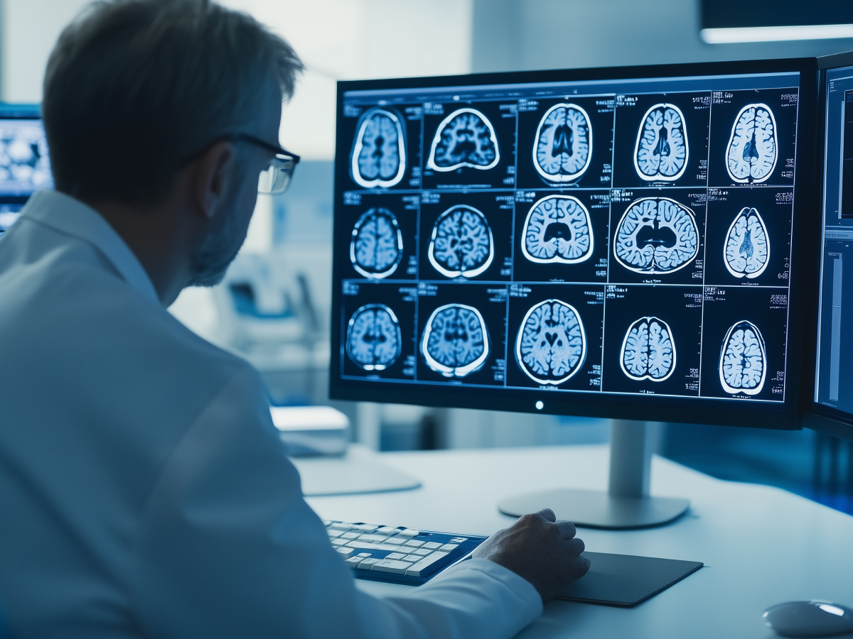
Breakthrough in MRI analysis with 3D models
GE Healthcare has stepped up and introduced the world’s first full-body 3D MRI foundation model (FM). This is a leap forward in how we interpret and use MRI data. Traditional MRI models sliced images into 2D segments. The problem is that you lose the bigger picture. And in healthcare, that “bigger picture” often determines life-altering decisions.
Now, this 3D FM changes everything. It lets us analyze the human body with an unprecedented level of precision. We’re talking about applications that span brain tumors, skeletal disorders, and cardiovascular diseases, some of the most challenging areas in medicine. Built on AWS infrastructure, the model was trained on a whopping 173,000 images from over 19,000 studies.
The potential impact here is immense. For procedures like biopsies, radiation therapy, and even robotic surgeries, this FM brings sharper insights and faster diagnoses. It’s achieved a 30% accuracy rate in matching MRI scans with text descriptions, a giant leap from the previous 3% rate.
Cost and computational efficiency in model development
When you’re working with data at this scale, massive 3D images that could crush most systems, you need to innovate, or you’ll hit a wall. GE Healthcare tackled this head-on by using AWS tools like Amazon SageMaker.
Using Nvidia A100 cores, they distributed the training across multiple GPUs. Which is a big deal because processing these enormous datasets on a single GPU will lead to rapid burnouts. SageMaker provided the distributed horsepower needed, cutting computational requirements by a staggering fivefold.
Cost efficiency was another key focus. With AWS elastic compute and storage solutions, GE Healthcare optimized resources. They moved infrequently accessed data to lower-cost tiers, giving financial feasibility. This means smaller hospitals and budget-constrained systems can access this game-changing technology without breaking the bank.
Introduction of multimodal and self-supervised learning techniques
Here’s where things get really futuristic. The 3D MRI FM integrates multimodal capabilities, linking images to text and classifying diseases with ease. It’s a unification of previously disjointed workflows, and it’s powerful.
Semi-supervised learning, especially the “student-teacher” approach, makes this model even more versatile. It doesn’t rely solely on large, annotated datasets, something that’s often a bottleneck in AI training. Instead, it learns from both labeled and unlabeled data, creating a scalable framework that hospitals with older machines and fewer resources can also benefit from.
This adaptability is key. GE taught the model to skip gaps and focus on what’s available, delivering accurate insights without getting bogged down.
Potential for medical applications
Now, let’s zoom out. While the model currently focuses on MRIs, its implications stretch far beyond. GE Healthcare is paving the way for applications in radiation therapy and x-ray imaging.
One of the most promising areas is in reducing manual effort. For example, radiologists spend hours marking organs during radiotherapy. The FM can automate this process, saving time and improving accuracy. It can also cut scan times for patients, making procedures more efficient and comfortable.
This change from single-disease models to a unified, scalable approach is huge. It leads to faster adaptation to new medical insights and broader applications across healthcare. Since 2020, GE’s AIR Recon DL feature has scanned 34 million patients. That’s a testament to the real-world impact.
Key takeaways
GE Healthcare’s 3D MRI FM is the next frontier in medical imaging. In combining precision, cost efficiency, and real-time capabilities, this innovation is set to massively improve patient care. Whether you’re looking at faster diagnoses or broader applications, the implications are enormous.


